Dr. Francesco Mintrone

N1™ Base

Anterior case

Dr. Francesco Mintrone

Italy

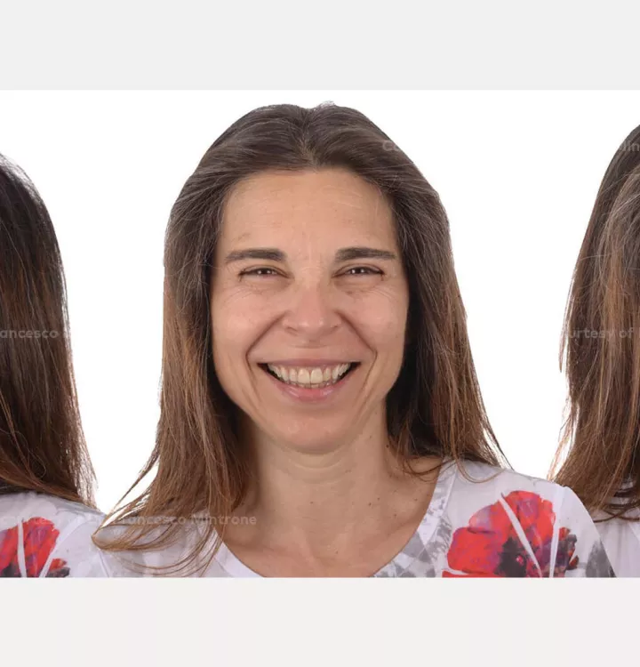
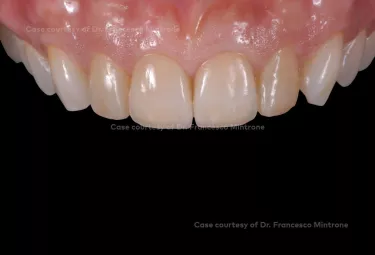
The request of the patient was to replace the fractured tooth and improve her smile, while minimizing the costs. Before starting surgical procedures, we decided to take care of the oral hygiene and periodontal situation. This enabled us to work in a healthy environment at the time of tooth extraction and implant placement. Immediate loading was performed on 21 with a socket shield technique. After the osseointegration of the implant, we decided to proceed with the digital approach to place the final restoration on the implant and a veneer on the other central incisor.
Case courtesy of Dr. Mintrone

Patient: 51-year-old female, poor oral hygiene, abrasion of the anterior teeth.

Clinical situation: impacted teeth 18, 48, 38. Deep fracture 21.

Surgical solution: extraction and implant placement on 21. 

Restorative solution: veneers on 11. Crown on implant on 21.

Surgical treatment: January 18, 2021

Rehabilitation delivery: June 7, 2021


Case courtesy of Dr. Francesco Mintrone.

Some products may not be regulatory cleared/released for sale in all markets.

Please use landscape mode when viewing this page on mobile.

Case courtesy of Dr. Mintrone

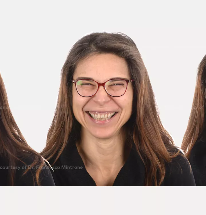
Esthetic analysis


Case courtesy of Dr. Mintrone

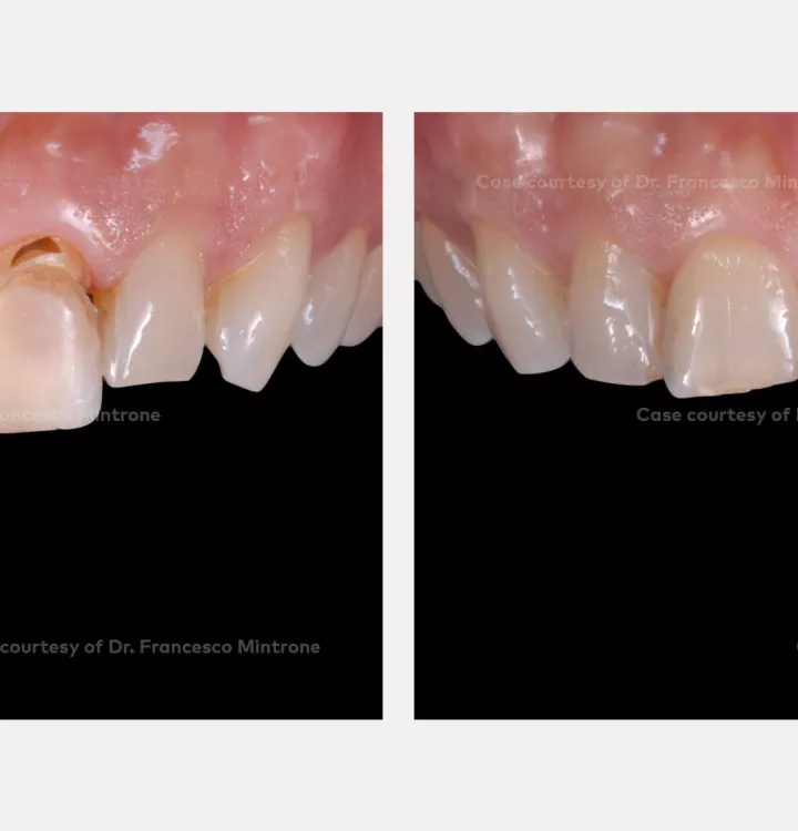
Initial clinical solution

Deep fracture of 21 and severe abrasion of 11.

Case courtesy of Dr. Mintrone

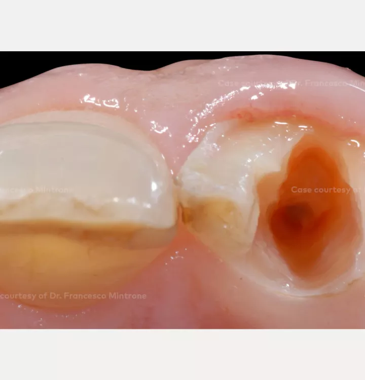
Initial clinical situation

Tooth residue of 21.

Case courtesy of Dr. Mintrone

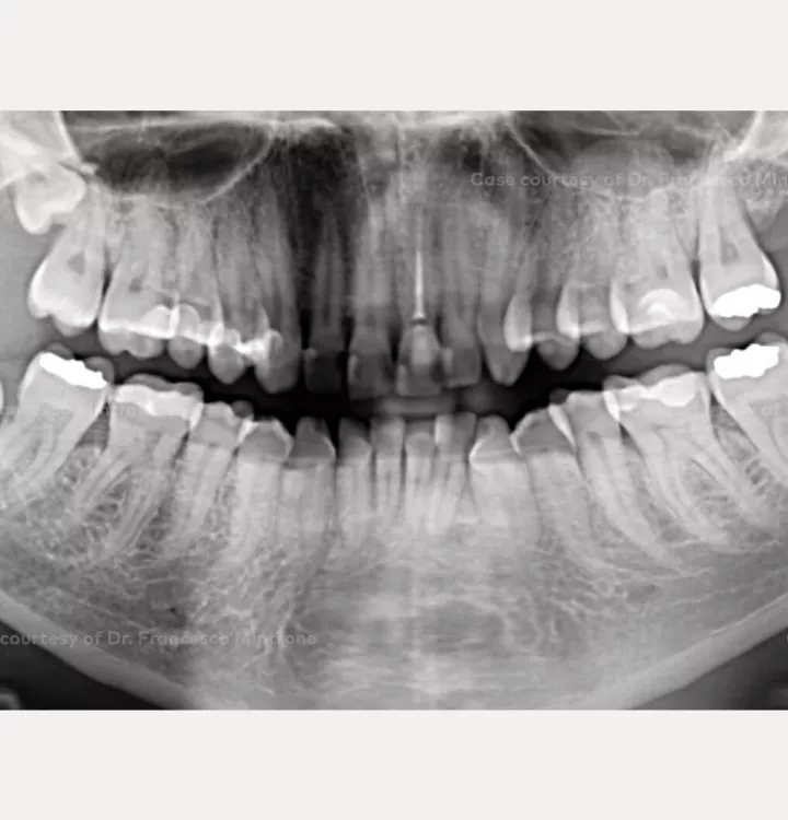
Initial clinical situation

Preoperative orthopantomagram. Incongruous fillings on all four maxillary incisors and impacted teeth 18, 38, and 48. Not visible: periapical lesions at tooth 21.

Case courtesy of Dr. Mintrone

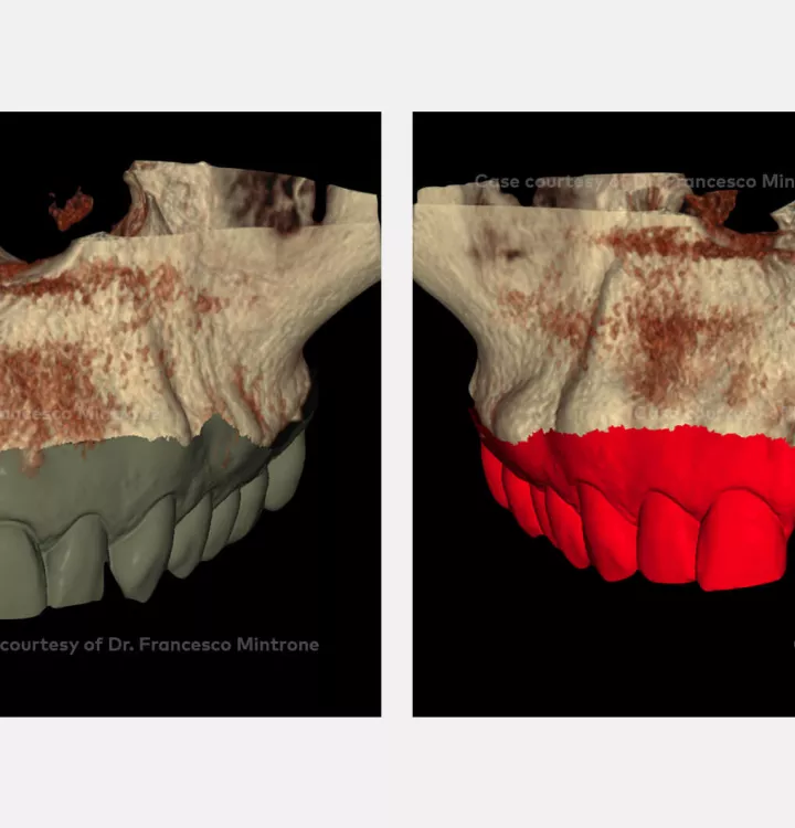
Treatment planning

Evaluation of the case with DTX Studio™ suite software.

Case courtesy of Dr. Mintrone

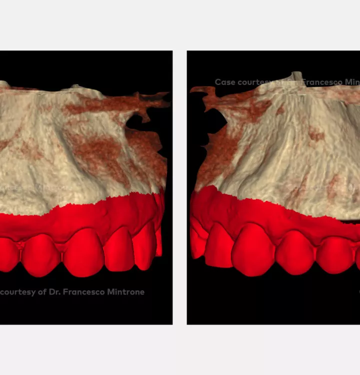
Treatment planning

Evaluation of the case with DTX Studio™ suite software.

Case courtesy of Dr. Mintrone

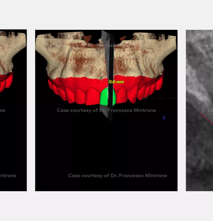
Treatment planning

Evaluation of the ideal position of the free gingival margin including crestal bone level assessment.

Case courtesy of Dr. Mintrone

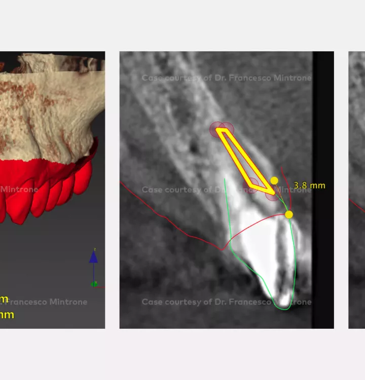
Treatment planning

Evaluation of the socket shield approach to preserve the buccal bone volume.

Case courtesy of Dr. Mintrone

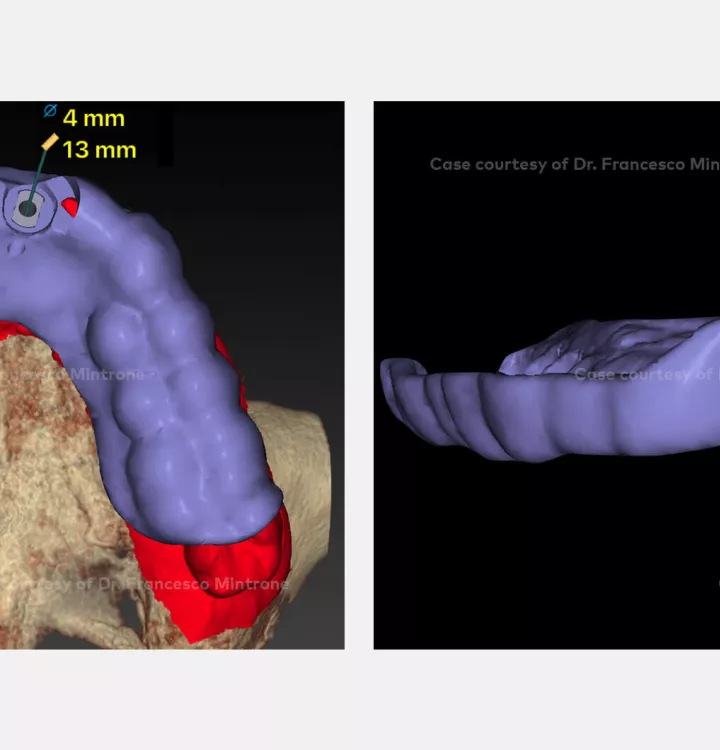
Treatment planning

Surgical guide for the 2 mm drill.

Case courtesy of Dr. Mintrone

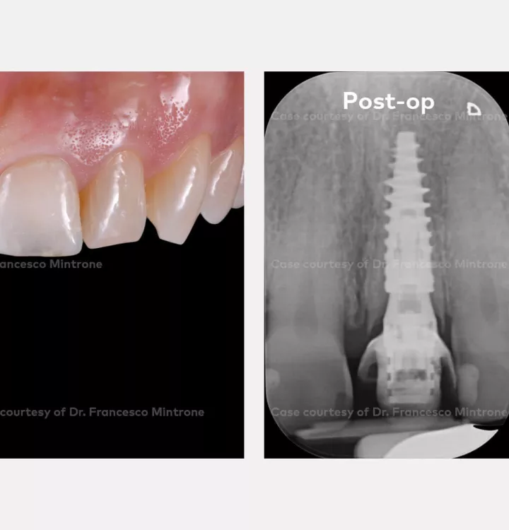
Surgical procedure

Provisional prosthesis and soft tissue healing after 90 days (left).

Peri-apical X-ray where the N1 Base and bone stability are visible (right).

Case courtesy of Dr. Mintrone

Restorative procedure

Mock-up to evaluate the prosthetic profile of the future rehabilitation.

Case courtesy of Dr. Mintrone

Restorative procedure

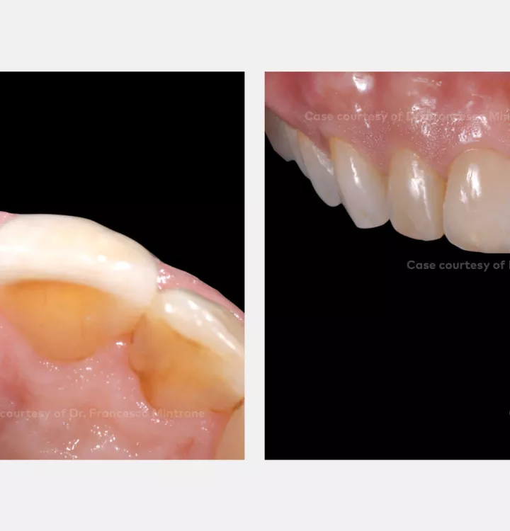
Minimally invasive preparation of the veneer on 11, and intraoral scanning.

Case courtesy of Dr. Mintrone

Restorative procedure

Intraoral scan.

Case courtesy of Dr. Mintrone

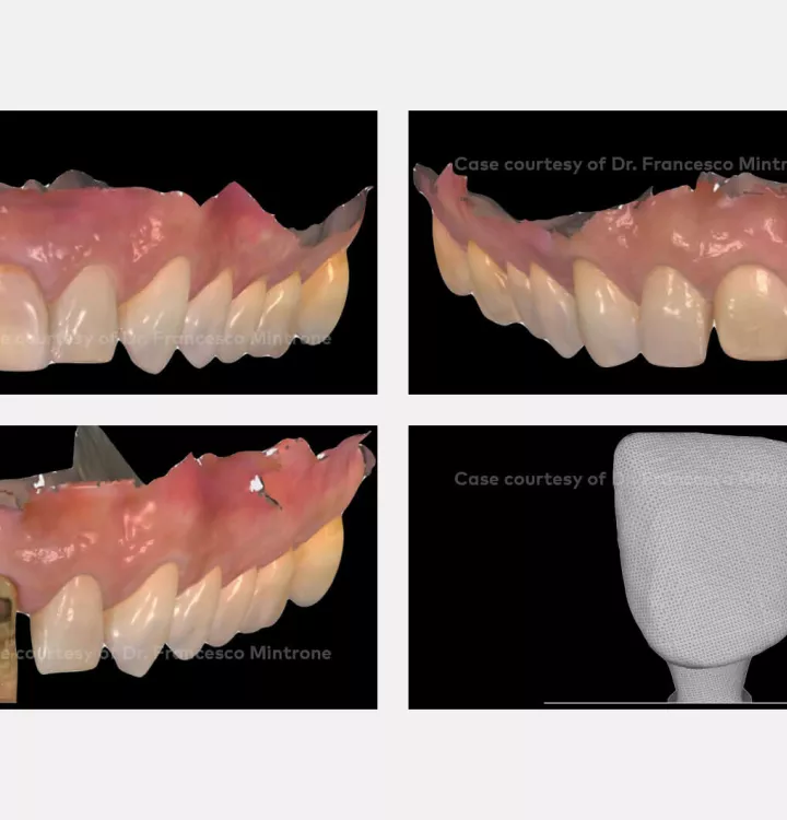
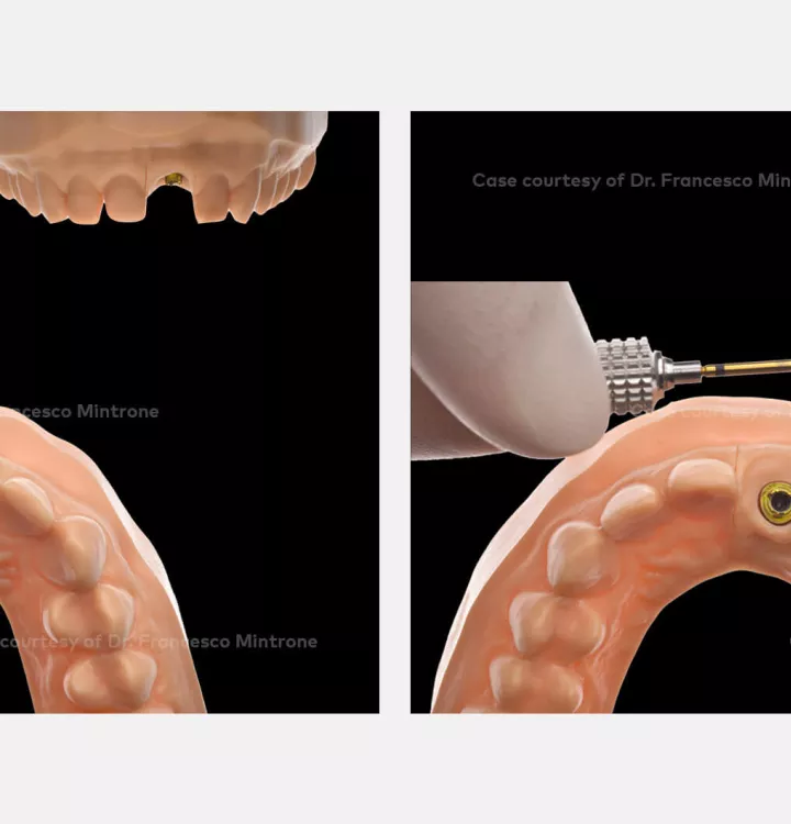
Laboratory procedure

Digital approach.

Case courtesy of Dr. Mintrone

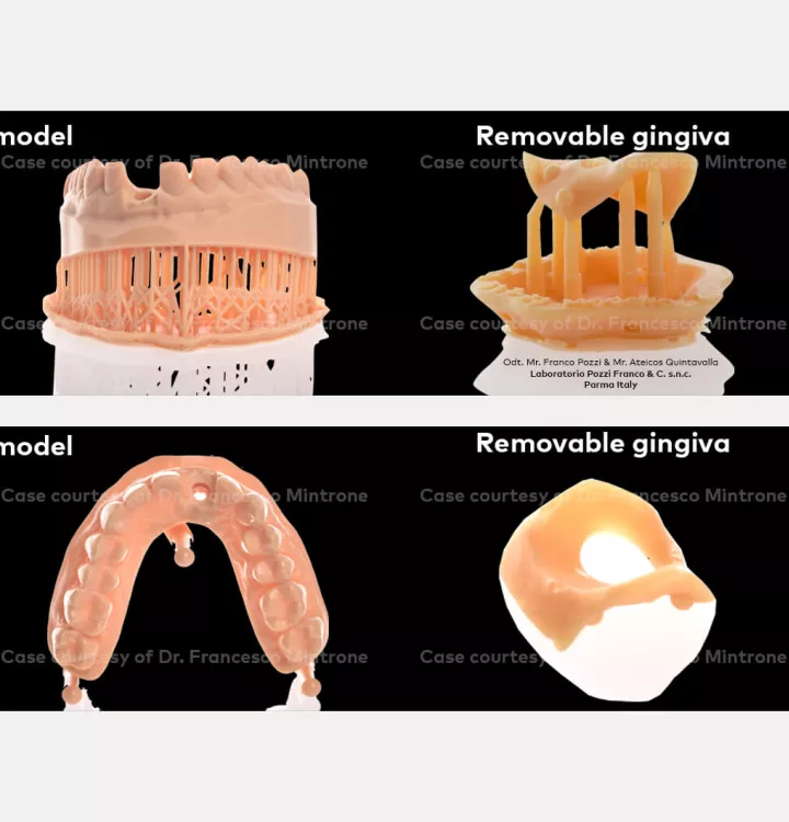
Laboratory procedure

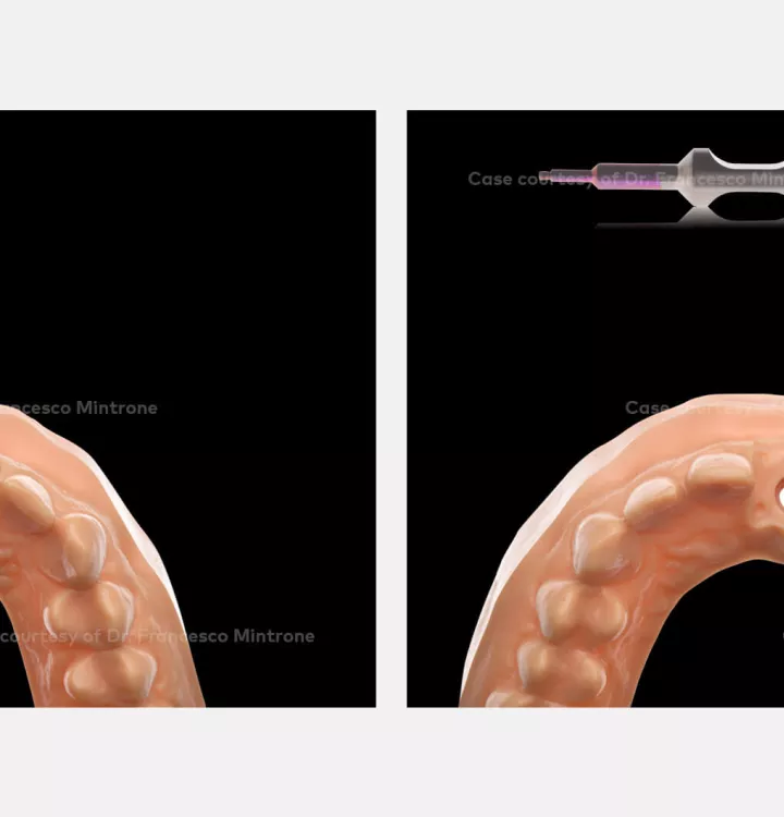
Digital approach (left).

Insertion tool for IOS implant replica Nobel Biocare N1 NP and RP (right).

Case courtesy of Dr. Mintrone

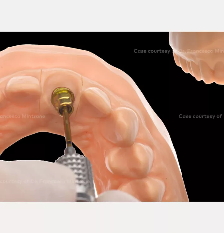
Laboratory procedure

Insertion tool for IOS implant replica Nobel Biocare N1 NP and RP.

Case courtesy of Dr. Mintrone

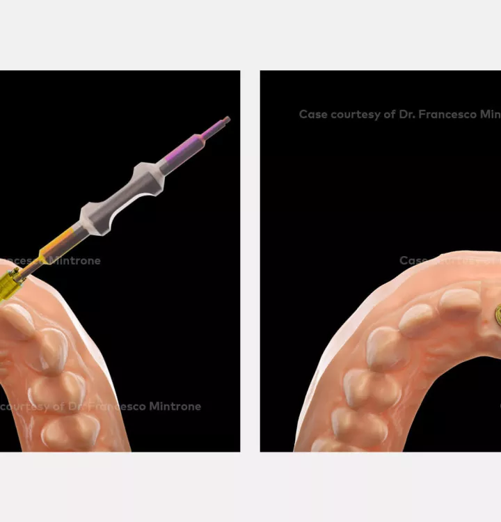
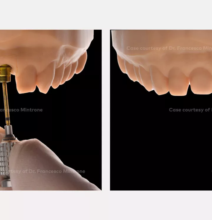
Laboratory procedure

Insertion tool for IOS implant replica Nobel Biocare N1 NP and RP.

Case courtesy of Dr. Mintrone

Laboratory procedure

Insertion tool for IOS implant replica Nobel Biocare N1 NP and RP.

Case courtesy of Dr. Mintrone

Laboratory procedure

Insertion tool for IOS implant replica Nobel Biocare N1 NP and RP.

Case courtesy of Dr. Mintrone

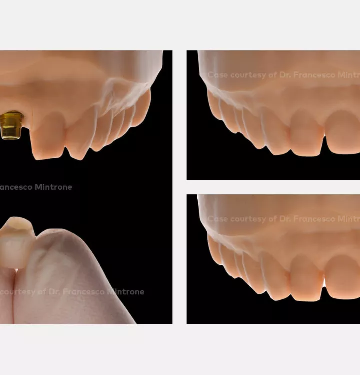
Laboratory procedure

Zirconia stamp to simplify the esthetic result.

Case courtesy of Dr. Mintrone

Laboratory procedure

Double approach with a zirconia stamp plus veneers cemented on top to simplify the esthetic result.

Case courtesy of Dr. Mintrone

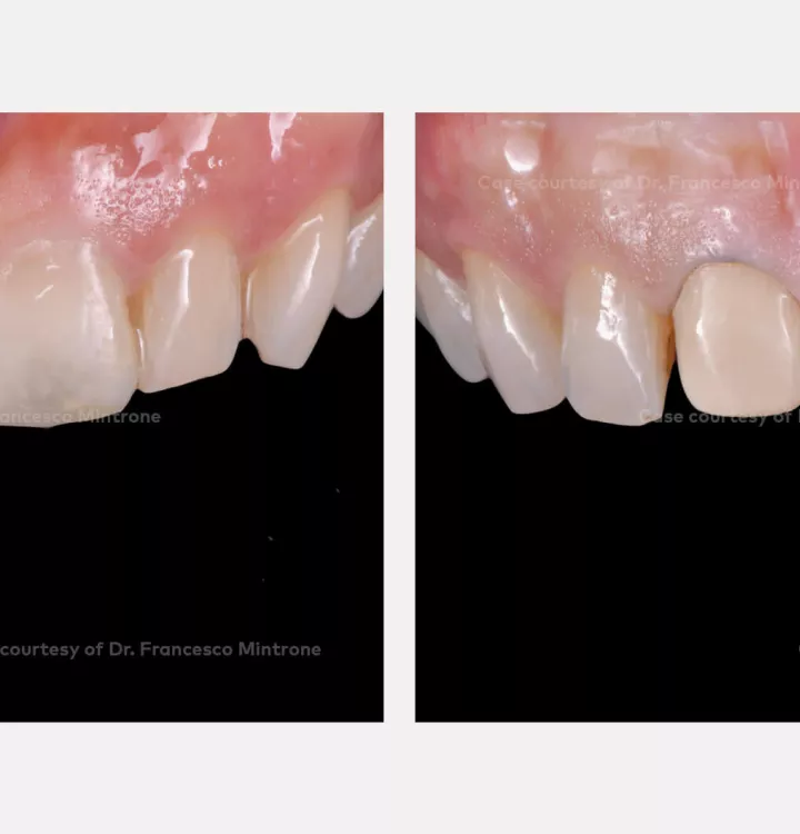
Outcome

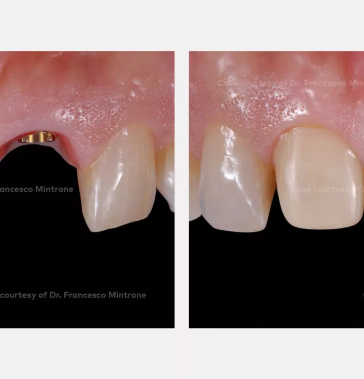
Good soft tissue stability around N1 base before the final delivery (left).

Good esthetic integration of the final rehabilitation (right).

Case courtesy of Dr. Mintrone

Outcome

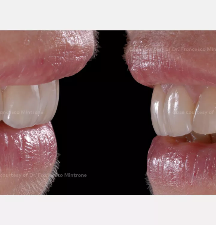
Good esthetic integration of the final rehabilitation.

Case courtesy of Dr. Mintrone

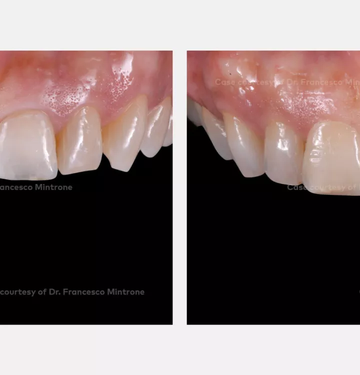
Outcome

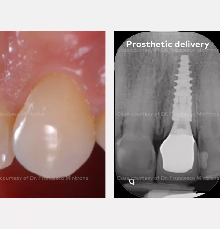
Soft tissue stability (left).

Peri-apical X-ray (right).

Case courtesy of Dr. Mintrone

Outcome

Good esthetic integration of the final rehabilitation.

Related products

Sign up for our blog update

Get the latest clinical cases, industry news, product information and more.

© Nobel Biocare Services AG, 2022. All rights reserved. Disclaimer: Nobel Biocare, the Nobel Biocare logotype and all other trademarks are, if nothing else is stated or is evident from the context in a certain case, trademarks of Nobel Biocare. Please refer to nobelbiocare.com/trademarks for more information. Product images are not necessarily to scale. All product images are for illustration purposes only and may not be an exact representation of the product. Disclaimer: Some products may not be regulatory cleared/released for sale in all markets. Please contact the local Nobel Biocare sales office for current product assortment and availability. For prescription use only. Caution: Federal (United States) law restricts this device to sale by or on the order of a licensed clinician, medical professional or physician. See Instructions For Use for full prescribing information, including indications, contraindications, warnings and precautions. Nobel Biocare does not take any liability for any injury or damage to any person or property arising from the use of this clinical case. This clinical case is not intended to recommend any measures, techniques, procedures or products, or give advice, and is not a substitute for medical training or your own clinical judgement as a healthcare professional. Viewers should never disregard professional medical advice or delay seeking medical treatment because of something they have seen in this clinical case.

Not all medical devices used during surgery are Nobel Biocare devices. Nobel Biocare recommends that manufacturer's IFUs are followed. Full procedure is not shown. Certain sequences have been cut. Please refer to applicable IFUs for full instructions.