Prof. Alessandro Pozzi

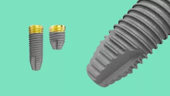
NobelParallel™ Conical Connection TiUltra™ Implant & X-Guide® for immediate loading of single implants in the esthetic zone in one visit

Dr. Alessandro Pozzi

Italy
Case courtesy of Dr. Alessandro Pozzi

Patient: 40-years-old female, no appreciable disease.

Clinical situation: Failed PFM crown on upper central incisor right side, with mobility, periapical infection and radiolucency, and oral fistula on the buccal side.

Surgical solution: Dynamic navigation and immediate loading of NobelParallel CC TiUltra in the esthetic zone in one visit.

Restorative solution: Immediate temporary prosthesis screw-retained porcelain fused to NobelProcera® ASC zirconia abutment.

Surgery date: April 2019.

Total treatment time: 6 months.

Tooth position: 11.


Case courtesy of Dr. Alessandro Pozzi.

Some products may not be regulatory cleared/released for sale in all markets.

Please use landscape mode when viewing this page on mobile.

Initial clinical situation

Failing PFM crown on tooth 11. Gingival recession, vertical super-eruption of the tooth misalignment of the gingival contour compared to tooth 21, inversion of the zenith position, buccal fistula. Challenging case for immediate loading due to malocclusion including a deep bite and poor canine guidance on the right side.

Treatment planning

Dynamic navigation for immediate tooth replacement in a single visit: CBCT and IOS scanning, DTX Studio™ implant 3D planning, DTX X-Guide streamlined execution.

Preoperative clinical frontal view and periapical X-ray. Failed PFM crown on upper central incisor right side with a clearly visible fistula on the buccal side and periapical infection and radiolucency.

Treatment planning

DTX Studio Implant 3D planning and rendering, and cross section snapped to the implant axis. The implant is positioned to bypass the radiolucency and engage the maximum quantity of native bone to guarantee sufficient primary stability. The angulation was designed to deliver a screw-retained immediate temporary crown.

Surgical procedure

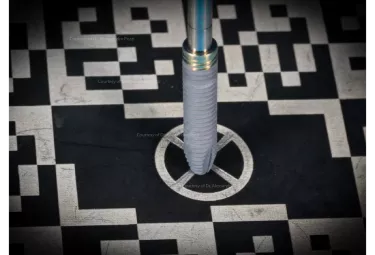
X-Guide Dynamic Virtual Guidance with 360° of control of implant drilling angulation and position.

Surgical procedure

Minimally invasive tooth extraction with a flapless approach, removal of the infected periapical tissues, and a careful debridement of the postextractive socket, followed by X-Guide-mediated implant placement, autogenous cortical plate graft to seal the buccal plate perforation, and augmentation with xenogeneic soft tissue matrix to enhance gingival architecture healing and thickening.

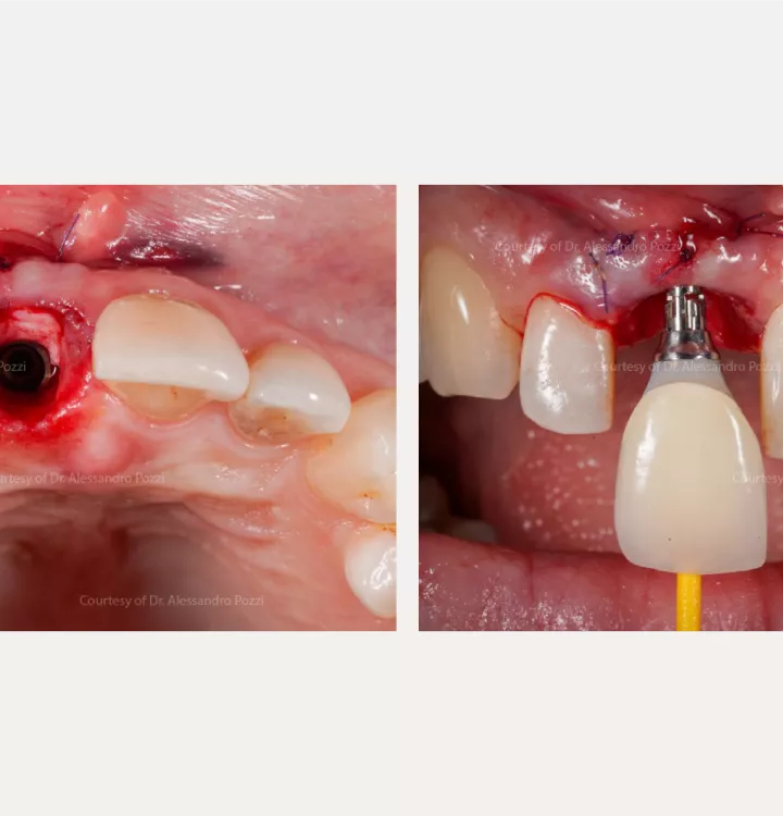
X-Guide-mediated implant positioning according to the prosthetically- and surgically-driven coordinates planned in the DTX Studio Implant software (left).

NobelParallel Conical Connection TiUltra NP 3.75 x 18 mm in the postextraction socket placed to allow the housing of the xenogeneic soft tissue matrix (right).

Surgical procedure

Intraoperative occlusal view after grafting on the buccal side. The xenogeneic collagen matrix packed and secured on the buccal side and interproximally (left).

A screw-retained immediate temporary crown on the Temporary Snap Abutment delivered on the day of surgery (right).

Restorative procedure

Postoperative view after 2 weeks: the minimally invasive X-Guide driven surgical positioning allowed fast healing and integration of the soft tissue matrix and bone tissue graft.

Restorative procedure

Case courtesy of Dr. Alessandro Pozzi

Frontal view of the definitive screw-retained porcelain fused to NobelProcera ASC zirconia abutment. 3D printed definitive mastercast with removable gum (left).

Palatal view of the definitive screw-retained porcelain fused to NobelProcera ASC zirconia abutment (right).

Outcome

Natural-looking appearance with continuous improvement of soft tissue outcomes.

Frontal view of the definitive screw-retained porcelain fused to NobelProcera ASC zirconia abutment. 1-year follow-up (top left).

Frontal view of the definitive screw-retained porcelain fused to NobelProcera ASC zirconia abutment. 2-year follow-up (bottom left).

1-year follow-up and 2-year follow-up. Periapical X-ray (right).

Related products

Sign up for our blog update

Get the latest clinical cases, industry news, product information and more.

© Nobel Biocare Services AG, 2021. All rights reserved. Nobel Biocare, the Nobel Biocare logotype and all other trademarks are, if nothing else is stated or is evident from the context in a certain case, trademarks of Nobel Biocare. Please refer to nobelbiocare.com/trademarks for more information. Product images are not necessarily to scale. All product images are for illustration purposes only and may not be an exact representation of the product. Some products may not be regulatory cleared/released for sale in all markets. Please contact the local Nobel Biocare sales office for current product assortment and availability. Caution: Federal (United States) law or the law in your jurisdiction may restrict this device to sale by or on the order of a dentist or a physician. See Instructions For Use for full prescribing information, including indications, contraindications, warnings and precaution. Dr. Alessandro Pozzi is a paid consultant for Nobel Biocare. The opinions expressed are those of the doctor. Nobel Biocare is a medical device manufacturer and does not dispense medical advice. Clinicians should use their own professional judgment in treating their patients. Individual patient results may vary.